Electron microscopy (EM) Laboratory
Electron microscopy (EM) Laboratory
The scanning electron microscope (SEM) can be applied to study mineral surfaces and complex textures in geological materials from micron to cm scale and also acquire information on the chemical composition.
Electron microscopy is applied in investigations of mineralogy, mineral composition, petrology and morphology. It has relevance for basic research as well as applied research within the mineral industry, construction materials and environment. SEM can for example be used for detection of asbestos particles in and problematic clay minerals in oil reservoirs.
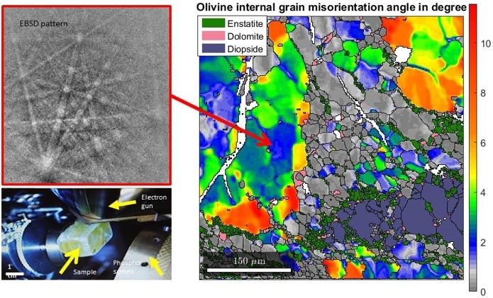
EBSD enables in-situ measurement of mineral orientation, making it possible to study grain structures and deformation mechanisms as well as growth processes, including topotaxial relations.
Cathodoluminescene can be used to image defect structures of minerals, which can be associated with grain growth and crack healing.
PTA is used to quantify mineralogy and mineral associations and to characterise how minerals occurs in crushed samples. Mineral liberation analyses is one of the main tasks for PTA.
The department collaborates with the electronmicroscopy lab at NTNU. The collaboration concerns three electron microscopes:
- Hitachi SU-6600 with CL, BED, 2x EDS, EBSD.
- Zeiss SUPRA 55VP (low vacuum, BED, EBSD, EDS).
- Zeiss ULTRA 55 (BED, EDS, EBSD) and also electron microprobe for quantitative mineral chemistry (5WDS, EDS, CL, BEI).
For more details on the EM lab, please visit Electron Microscopy Lab in Materials Science at NTNU.
Laboratories at IGP
- Chemical/Mineralogical Laboratory
- Petrographic- and Ore Microscopy Laboratory
- Electron microscopy (EM) Laboratory
- Mineral processing Laboratory
- Thin Section Laboratory
- Geological Engineering Laboratory
- Rock Mechanics Laboratory
- Rock Magnetic Laboratory
- Hall laboratory
- Reservoar Laboratory
- Schlumberger geolab
Workshops: