Medical imaging
Medical Imaging

Wireless capsule endoscopy
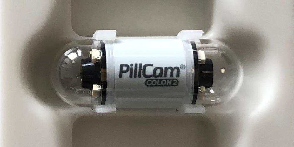
Imagine swallowing a tiny camera that can travel through your digestive system and take pictures of your intestines. This is not science fiction, but a real medical technique called wireless capsule endoscopy (WCE). WCE can help doctors diagnose various diseases and conditions, such as inflammation, bleeding, and cancer, in a minimally invasive way (Floor et al., 2020).
But WCE is not without challenges. The camera produces a huge amount of data, which can be hard to analyse and store. The image quality can be low due to motion blur, illumination changes, and capsule rotation, and the annotation of the images can be limited by the availability and expertise of the doctors.
This is where the Colourlab comes in. Our researchers have been working on developing novel methods and algorithms for WCE image analysis and processing, in collaboration with hospitals and medical professionals.
3D reconstruction
One of our achievements is a method for creating a 3D model of the human colon from WCE video. This can help doctors better visualize and understand the colon anatomy and pathology. In Floor et al. (2022), they used a SLAM-based approach to estimate the capsule orientation and position, and a structure from motion-based approach to refine the resulting 3D point cloud. They showed that their method can produce accurate 3D reconstructions.
Artificial Intelligence
Another achievement comes by weaving simple prior knowledge into how deep networks learn to distinguish pathologies, resulting in improving the capacity of neural networks in distinguishing between different bowel pathologies compared to other approaches (2021). Vats et al. (2022a) also used a generative adversarial network (GAN) based network to learn the distribution of WCE data for realistic simulation of different pathologies.
Vats et al. (2022b) have also introduced a multichannel residual cues network for fine-grained classification of VCE images. This means that they can identify subtle differences between different types of abnormalities, such as polyps, ulcers, and tumors. The complementary information from different colour channels and residual cues were exploited to enhance the feature representation and discrimination of the VCE images. Their network was tested on a large-scale WCE dataset and showed that it can beat state-of-the-art methods for fine-grained classification (Vats et al., 2022b).
In addition, Floor et al. (2020) developed a post-processing method for reducing the errors in WCE video caused by noise. They used a combination of temporal and spatial inpainting to improve the image quality of the WCE video. Their method was evaluated on real WCE data and showed that it can significantly reduce the errors and artifacts.
These are just some examples of Colourlab’s research on medical imaging, especially WCE. They show the potential of Colourlab’s methods and solutions for advancing the field of medical imaging and improving the clinical outcomes for patients. Further studies are being conducted, and you can read more about the research in the below mentioned publications.
This text is partially generated by Microsoft Copilot (AI).
