Laboratories - CEMIR
Laboratory facilities and infrastructure
The CEMIR laboratories are located in the Knowledge Centre and the Gastro Centre at the Øya Campus of St.Olavs Hospital and NTNU.
New laboratories opened in the Knowledge Centre in June 2014, and in October 2015 we opened a new Biosafety Level Three laboratory (BSL-3), offering the highest level of security for research on viruses and bacteria in Norway. CEMIR hosts first-class laboratories with state-of-the-art equipment for performing research on cells, tissues and microorganisms.
State-of-the-art equipment
-
High resolution STED confocal microscope
-
Total internal reflection fluorescence (TIRF) microscope
-
Live cell- and spinning disk confocal microscopes
-
Image flow cytometer
-
Cell sorter
-
A confocal microscope installed in a biosafety level 3 facility
The BSL-3 lab contains an advanced Leica SP8 confocal microscope making it possible to study infections in immune cells with viable Mycobacterium tuberculosis and HIV virus.
Imaging Core Facility
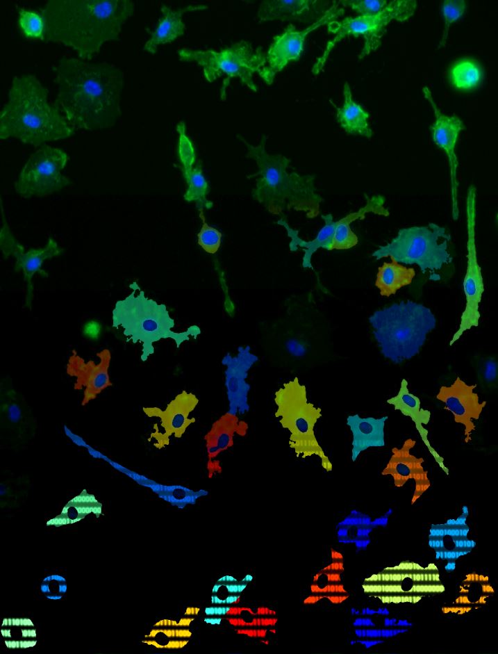
Researchers and students at CEMIR have access to a multitude of different imaging techniques, for live cell studies as well as imaging of fixed cells and tissue preparations.
These instruments are all part of the Cellular and Molecular Imaging Core Facility (CMIC) at Faculty of Medicine, NTNU. The CEMIR director is the scientific leader for this core facility. CMIC is closely integrated with the CEMIR’s laboratories and its research groups, and CEMIR scientists are the biggest user group of CMIC. The core facility offers several services: standard confocal microscopy and live cell imaging, fully automated high-throughput image acquisition, image visualization and image processing, immune histochemistry and electron microscopy.
In 2014 a super resolution light microscope and a total internal reflection fluorescence microscope were installed at CMIC and placed in the CEMIR laboratories. In 2016 CMIC purchased a Pico Quant single molecule detection (SMD) upgrade for our Leica SP8 STED 3X super-resolution microscope. This add-on is in particular useful for studying molecular interactions in cells. In addition, CEMIR researchers are using Focused-Ion Beam Scanning Electron Microscopy (FIB-SEM) in collaboration with the NTNU Nanolab. A new 3-D serial block face scanning electron microscope has also recently been added to the instrument park.