Summer school speakers
AI in Neuroscience summer school speakers and contributors
Jon Sporring
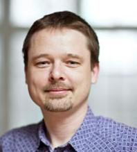
Professor, Dept. of Computer Science, Copenhagen University
Title: “Stochastic geometry in Bioimaging”
Abstract:
Geometrical measurements of biological objects form the basis of many quantitative analyses. Hausdorff measures such as the volume and the area of objects are simple and popular descriptors of individual objects; however, for most biological processes, the interaction between objects cannot be ignored, and the shape and function of neighboring objects are mutually influential. In this paper, we present a theory on the geometrical interaction between objects inspired by K-functions for spatial point-processes. Our theory describes the relation between two objects: a reference and an observed object. We generate the r-parallel sets of the reference object, calculate the intersection between the r-parallel sets and the observed object, and define measures on these intersections. The measures are simple, like the volume or surface area, but describe further details about the shape of individual objects and their pairwise geometrical relation. Finally, we propose a summary-statistics. To evaluate these measures, we present a new segmentation of cell membrane, mitochondria, synapses, vesicles, and endoplasmic reticulum in a publicly available FIB-SEM 3D brain tissue data set and use our proposed method to analyze key biological structures herein.”
Bio:
Jon Sporring received his Master and Ph.D. degree from the Department of Computer Science, University of Copenhagen, Denmark in 1995 and 1998, respectively. Part of his Ph.D. program was carried out at IBM Research Center, Almaden, California, USA. Following his Ph.D, he worked as a visiting researcher at the Computer Vision and Robotics Lab at Foundation for Research & Technology - Hellas, Greece, and as an assistant research professor at 3D-Lab, School of Dentistry, University of Copenhagen. Since 2003 he has been employed as an associate professor at the Department of Computer Science, University of Copenhagen. From 2008-2009 he was a part-time Senior Researcher at Nordic Bioscience a/s. In the period 2012-13, he is a visiting professor at the School of Computer Science, McGill University, Montreal, Canada. Jon Sporring also co-founded DigiCorpus Aps in 2012 and served as Chief research officer of the company from 2012-16 developing computer vision-based systems for automatic feedback for physiotherapeutic rehabilitation. In 2007-2012, 2015-2019, and 2021 he was Deputy head of the Department for Research at the Department of Computer Science, University of Copenhagen. Since 2018, he is a full professor at the University of Copenhagen, and since 2019 he is the Deputy head of the Center for Quantifying Images for MAXIV (QIM).
Jens Nyengaard
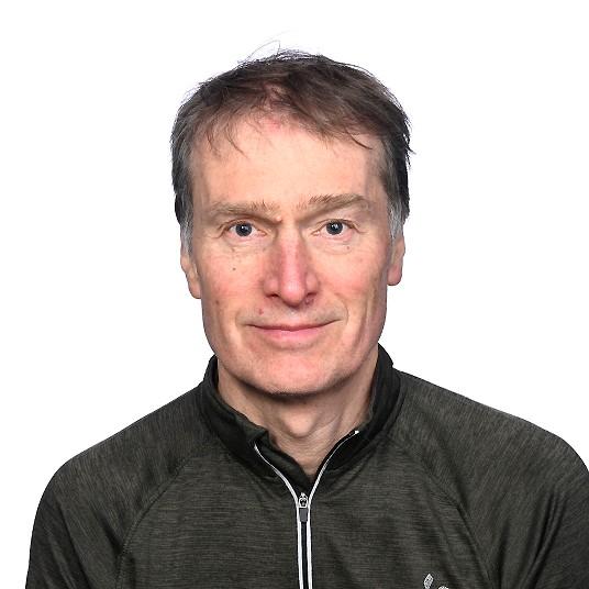
Neuroscientist, Professor, Dept. of Clinical Medicine, Aarhus University, Denmark
Title: “Characterizationof Pyramidal Cells in Layer III of Brodmann Area 46 in Schizophrenia and Depression”
Abstract:
Schizophrenia and depression are neuropsychiatric disorders that impair human thoughts, emotions, and behavior. Radio medical observations and histological studies have confirmed structural and functional alterations in brains from patients with schizophrenia and depression. Among these, abnormal activation in the dorsolateral prefrontal cortex, including Brodmann Area 46 (BA46) is prevalent. This irregular brain activity in BA46 could be a consequence of the number and spatial distribution of neurons as well as soma size and shape.
The primary objective of this Ph.D. dissertation is to examine and study the impact of neuroanatomical differences in layer III of BA46 in postmortem schizophrenic and depressive patients compared to healthy controls. We used autopsy human brain tissue from ten healthy subjects, ten subjects with schizophrenia, eleven suicide subjects with a history of depression, and eight people who had major depressive disorder (MDD) without committing suicide. Advanced methods using stochastic geometry and machine learning to reconstruct neurons in 3D were developed to characterize the neuronal organization, size, and shape. Besides, we also used stereology and estimated volume tensors to quantify the total volume in layer III of BA46, the total cell number, number density, cell soma volume, and shape of pyramidal cells.
Our findings showed that schizophrenic patients tend to have a smaller volume of layer III in BA46. Compared to healthy control and suicide groups, both schizophrenic and MDD patients show a reduction in cell number, density, and volume. There is no significant difference in the elongation index of pyramidal cells in the four groups, but there is a notable displacement of the nucleolus between the suicide and schizophrenic groups. No significant changes among groups are observed in terms of shape and orientation. We discovered that the positions of neurons are either arranged into columnar structures or have some repulsive behavior against each other. These results imply that the neurons are not randomly organized in 3D space but follow a columnar pattern. Furthermore, this project emphasizes and broadens the applicability of old archived autopsy-based material to examine cell quantities and their spatial distribution.
Bio:
Prof. Nyengaard has extensive experience in leading laboratories, scientific projects and personnel in both Denmark and abroad (USA, Switzerland, Germany, Saudi Arabia, China and Vietnam). The 391 publications have a high impact factor (H-index=63), they are widely cited (>21400 citations) and they cover a broad range of medical fields. His laboratory “Core Centre for Molecular Morphology” has a world-renowned profile in quantitative microscopy within medical research. This profile has in the last 20 years been extended into a more biomolecular direction within neuropsychiatry/neurology and translational medicine with specific molecular labelling techniques for microscopy including advanced sampling methods, deep learning, complex fluorescence methods (FRET, FLIM, FRAP, FCS) and different types of electron microscopy: immuno-transmission electron microscopy, cryo-transmission electron microscopy, focused ion beam scanning electron microscopy and serial block face scanning electron microscopy. The scientific goals and strategic portfolio of the Nyengaard lab encompass the discovery and translational research in human neuropathologies including novel solutions for Neuroscience community. The developed and applied quantitative methods are in use in many hundreds laboratories around the world and this achievement has stimulated many international, interdisciplinary initiatives and collaborations with both public and private partners.
Armin Iraji
Assistant Professor, Dept. of Computer Science, Georgia, Georgia State University, United States
Title of talk and tutorial: “Dynamic functional connectivity”
Abstract:
The analysis of functional magnetic resonance imaging (fMRI) data can greatly benefit from the utilization of flexible analytic approaches. Data-driven techniques enhance our ability to capture dynamic functional connectivity and uncover important relationships in the data, which, when ignored, can result in misleading information and imprecise inferences
In this presentation, I will introduce independent component analysis (ICA) as a powerful data-driven tool for estimating functional sources in fMRI data. Moreover, I will showcase multiple methods for estimating the spatial and temporal dynamics of the brain, highlighting their application in both investigations of brain health and disorders.
I will also provide a comprehensive guide to the openly accessible GIFT software, demonstrating its practical usage in estimating intrinsic connectivity networks (ICNs) and capturing dynamic states. Through hands-on examples, we will explore the application of independent component analysis (ICA) and dynamic functional network connectivity (dFNC) analyses. This demonstration will showcase the wide-ranging possibilities and exciting opportunities within the field, emphasizing the potential for advancements and breakthroughs.
Bio:
Armin Iraji is an Assistant Professor in the Department of Computer Science at Georgia State University and a full-time faculty member of the Tri-Institutional (Georgia State, Georgia Tech, Emory) Center for Translational Research in Neuroimaging and Data Science (TReNDS). Armin's research primarily focuses on obtaining a comprehensive mapping of brain functional connectivity and capturing its detailed inter- and intra-characterization. He develops data-driven techniques and mathematical models to estimate the spatiotemporal dynamics of the brain and dynamic alterations in various cognitive and mental states.
Tom Ryen

Head of Department, Department of Electrical Engineering and Computer Science, UiS
Bio:
Tom Ryen has since 2016 been Head of department at the Department of Electrical Engineering and Computer Science at the University of Stavanger (UiS). He has a PhD in digital signal processing. He has been an Associate Professor in computer science at UiS since 2002, with the exception of the year 2013, when he held a research position at SINTEF. In addition to the department head job, he was active in the establishment of Stavanger AI Lab, which is a virtual lab at UiS with a focus on research, innovation and education in artificial intelligence. Tom Ryen is Chair of the Board in NORA (Norwegian Artificial Intelligence Research Consortium).
Antorweep Chakravorty
Associate professor, SAIL, Stavanger AI Lab
Tentative title of talk: “Micro-gesture recognition and AI”
Trygve Eftestøl
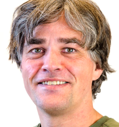
Professor, Dept. of electrical engineering and computer science, Biomedical Data Analysis laboratory and Cognitive laboratory
Title: Hands on workshop on feature extraction from biomedical signals
Abstract:
In this workshop we will take you through some basics of data handling and signal processing. You will go through the steps necessary to read data from a file into a data analysis framework (Scientific Python). Furthermore you will do some filtering to remove noise from the signal, transform the signal to the frequency domain, and finally compute features from both the time domain and the frequency domain. You will also visualise the features in scatter plots.
Bio:
Trygve Eftestøl was born in Bergen, Norway, in 1967. He received the B.Eng. degree in electrical engineering from Bergen College, Bergen, in 1992 and the M.Sc. degree in cybernetics and the Ph.D. degree in signal processing from Stavanger University College, Stavanger, Norway, in 1994 and 2000, respectively. He is currently a Professor at the Department of Electrical and Computer Engineering, University of Stavanger (UiS). He is deputy leader of the Biomedical Data Analysis Laboratory and in the steering comittee of the Cognitive and Behavioral Neuroscience Lab. Both groups are affiliated to UiS. Eftestøl served as an expert reviewer for the evidence evaluation process for the 2005 and 2010 International Consensus Conference on cardiopulmonary resuscitation and emergency cardiovascular care science with treatment recommendations appointed by the International Liaison Committee on Resuscitation, European Resuscitation Council and American Heart Association. Eftestøl also contributed as coauthor on a chapter on Analysis and Predictive Value of the Ventricular Fibrillation Waveform in the second edition of the book: Cardiac Arrest - The Science and Practice of Resuscitation Medicine published by Cambridge University Press in 2007. His main research interests include biomedical signal and image analysis, and machine learning applie within fields like out-of-hospital and newborn resuscitation, sports medicine, cognitive neuroscience, myocardial infarction, and digital pathology. He is a senior member of IEEE. He has published 90 papers in SCI-IF journals and over 100 contributions to scientific conferences.
Kjersti Engan
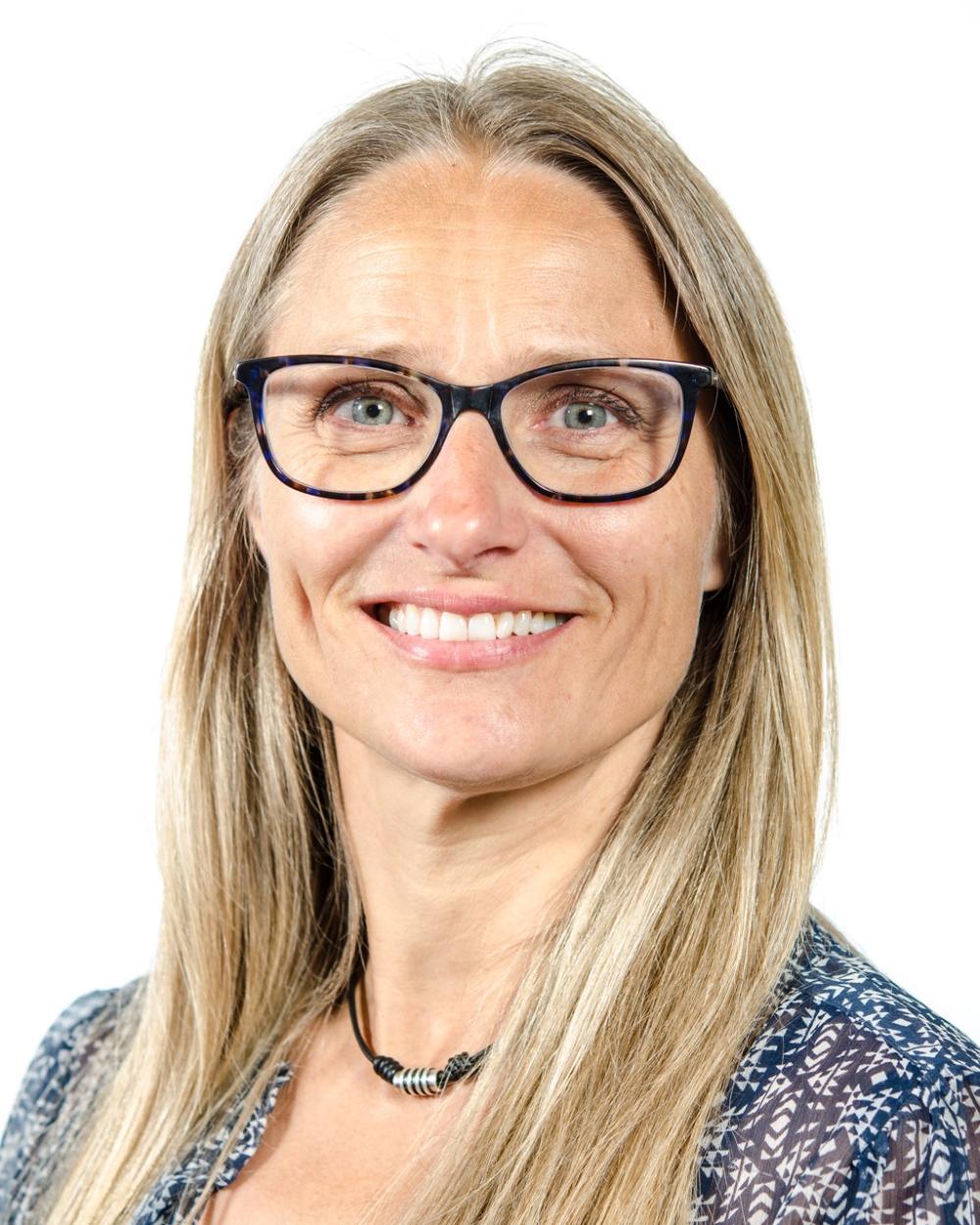
Professor, Dept. of electrical engineering and computer science, Biomedical Data Analysis laboratory
Title: "AI in Medicine - from digital pathology to newborn survival”
Abstract:
Artificial Intelligence (AI) has emerged as a powerful tool in various fields, and its potential in medicine is rapidly becoming evident. This talk explores some of the progress of AI applications in medicine, focusing on two areas: digital pathology and newborn survival. Digital pathology has changed the field of diagnostic pathology by enabling the digitization of histopathological images into Whole Slide Images (WSI). AI algorithms, combined with image processing, have demonstrated promising capabilities in segmentation of cancerous regions, visualization and diagnostic and prognostic predictions from WSI. In this talk we will look at some of the results from urinary bladder cancer and Melanoma (skin cancer) predictions. Globally 2 million newborn dies every year within the first 24 hours of life, and birth asphyxia is the main reason. Evidence based research on newborn resuscitation procedures are highly sought for. In this talk we will look at which data we can collect and how AI can help us learn. Particularly we will look at fetal heart rate monitoring and activity recognition from video during birth and resuscitation.
Bio:
Kjersti Engan is a professor at the Electrical Engineering and Computer Science department at the University of Stavanger (UiS), Norway. She received the BE degree in electrical engineering from Bergen University College in 1994 and the M.Sc. and Ph.D degrees in 1996 and 2000 respectively, in electrical engineering and information technology from the UiS. She is the leader of the Biomedical data analysis lab - BMDLab - at UiS. Her research interests are in signal and image processing and machine learning with emphasis on medical applications and in dictionary learning for sparse signal and image representation. She has a particular interest in AI for newborn survival, stroke detection from CTP imaging and AI in computational pathology. She is a senior member of IEEE and a member of NOBIM. She has served as Associate editor and Senior Area editor for IEEE Signal Processing Letters, as a member of IEEE Image, Video, and Multidimensional Signal Processing Technical Comittee (IVMSP), associate editor for SIAM Journal on Imaging Sciences (SIIMS), and as board member of NOBIM. She is currently on the board of NORA (Norwegian Artificial Intelligence Research Consortum).
Mahdieh Khanmohammadi
Associate professor, Dept. of electrical engineering and computer science, Biomedical Data Analysis laboratory
Title: Applying AI based techniques in different settings related to Neuroscience.
Abstract:
- Analyzing event-related potentials (ERP) using machine/deep learning
The Event-Related Potentials (ERPs) data from five distinct subject groups are analyzed and classified. In this project techniques, such as Random Forest, Gradient Boosting, K-Nearest Neighbor classifiers, and the EEGNet approach, are employed. The performance of the models is evaluated using accuracy, precision, recall, and F1-score metrics.
- Deep Learning Footprints on Synaptic Vesicle Detection Using Electron Microscopy Images
Behavioral stress has shown to strongly affect the density and distribution of the synaptic vesicles within medial prefrontal cortex. To study the distribution of the synaptic vesicles their location within the neuron is essential. We employed convolutional neural network and compressed sensing through serial section transmission electron microscopy images to automatically detect the vesicles’ centers. The framework of the proposed system has proven to be robust however, such framework requires large number of training examples with diversity to optimize its parameters.
Bio:
Mahdieh Khanmohammadi, an associate professor at the University of Stavanger, has made significant contributions to the field of medical image analysis. She was born and raised in Iran pursued her passion for science and technology by studying bioelectrical engineering as her bachelor and continued to master’s degree in the same field at the University of Tehran. Mahdieh obtained her second master’s degree in computational engineering from University of Halmstad in Sweden. Motivated by her master's research, Mahdieh pursued a Ph.D. program at the University of Copenhagen in Denmark. There, she joined the image processing group at the Computer Science Department, working on a project in collaboration with the neuroscientists at Aarhus University and the Statistical and Mathematical Science Department at Aalborg University. Her doctoral research involved analyzing the effect of behavioral stress on the brain, utilizing microscopic images of rat brains. Following the completion of her Ph.D., Mahdieh continued her academic journey as a postdoctoral researcher at the University of Stavanger. In collaboration with cardiologists at Stavanger University Hospital, she focused on analyzing angiographic data of the heart to estimate blood velocity.
Aleksandra Sevic

PhD fellow, Faculty of Health Sciences, Facilitator at cognitive laboratory
Title: “From Signals to Insights: Integrating Physiological Signals for Neuroscience Research”
Abstract:
In this workshop, the participants will get the chance to visit the Cognitive lab at the UiS. During the workshop, participants will experience practical demonstrations of lab equipment, showcasing the possibilities and complexities of integrating diverse biosensor modalities (EEG, EDA, ECG, eye tracking, and facial expressions analysis) in neuroscience research.
Bio:
Aleksandra Sevic is a psychologist and a PhD Candidate at the Faculty of Health Sciences at the UiS. iMotions certified for human behavior research.
Simone Grassini
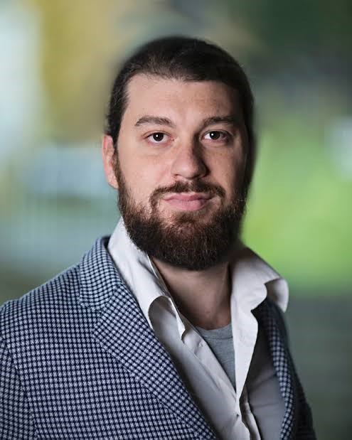
Associate professor, Faculty of Social Sciences
Title: "From Signals to Insights: Integrating Physiological Signals for Neuroscience Research" (joint session with Aleksandra Sevic)
Florenc Demrozi
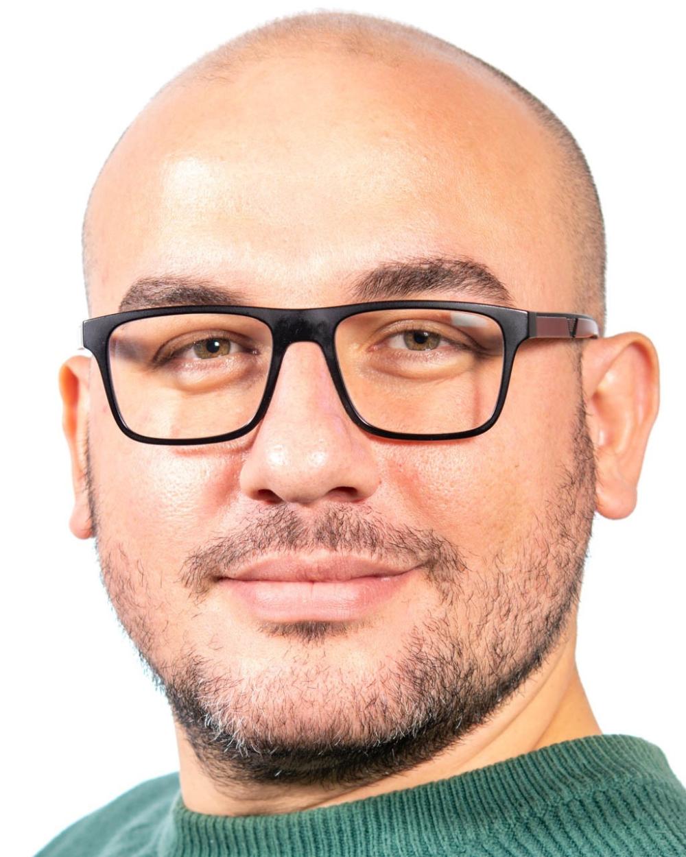
Associate professor, Dept. of electrical engineering and computer science, Biomedical Data Analysis laboratory;
Title: "The Synergy of Artificial Intelligence of Things (AIoT) and Sensor Technology in Human Activity Recognition (HAR) for Personalized Healthcare”
Abstract:
The integration of Artificial Intelligence of Things (AIoT) and sensor technology has opened up new horizons in the field of personalized healthcare. This talk explores the synergy between AIoT and sensor technology in the context of Human Activity Recognition (HAR) for personalized healthcare applications. HAR involves the identification and analysis of human activities to gain insights into an individual's health status, well-being, and overall lifestyle. By leveraging AIoT and sensor technology, HAR can be enhanced to provide more accurate, real-time, and personalized healthcare solutions.
Bio:
Florenc Demrozi currently holds the position of Associate Professor in Biomedical Engineering at the Department of Electrical Engineering and Computer Science, University of Stavanger in Norway. His research focuses on Artificial Intelligence of Things, Human Activity Recognition (HAR), Internet of Medical Things (IoMT), Sensors, and Measurements.
In May 2020, he completed his Ph.D. in Computer Science under the supervision of Prof. Graziano Pravadelli. His doctoral thesis titled "An IoT-based Virtual Coaching System for Assisting Activities of Daily Life" explored the development of a system that utilizes IoT technology to provide support for daily activities. Prior to that, he obtained his Master's degree (M.Sc.) in Computer Science and Engineering from the University of Verona in 2016, where his thesis focused on "Automatic generation of self-adaptive transactors from PSL assertions." He also earned his Bachelor's degree (B.Sc.) in Computer Science from the same university, under the guidance of Prof. Graziano Pravadelli. In April 2021, Florenc co-founded the IoT for Care (IoT4Care) research group at the Department of Computer Science, University of Verona, Italy. This research group is dedicated to developing Virtual Coaching Systems that utilize IoT infrastructure and the Extended Mind concept to assist individuals with special needs in their daily activities.
Florenc has also gained research experience as a visiting researcher at the University of Florida, Gainesville in 2019, and the Technical University of Chemnitz, Germany from 2020 to 2021. From January 2023 to March 2023, he received an AWS fellowship from the University of Limerick, Ireland, where he served as a visiting professor of Data Analytics for the new Immersive Software Engineering (ISE) B.Sc./M.Sc. degree program.
Ketil Oppedal
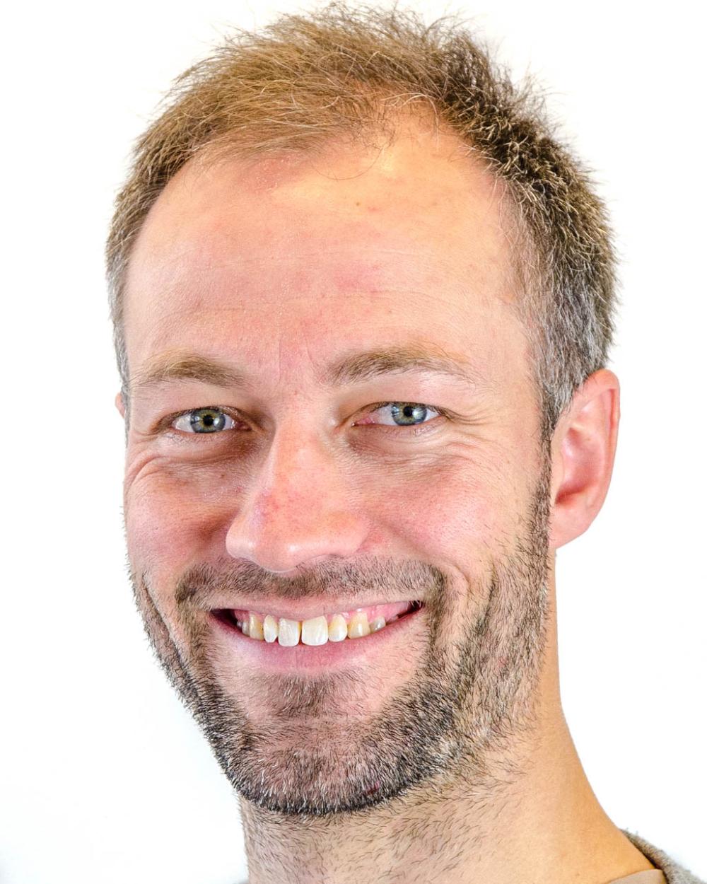
Associate professor, Dept. of electrical engineering and computer science, Biomedical Data Analysis laboratory
Title: “Inoduction to and applications of generative deep learning methods in medical imaging"
Abstract:
In this talk I want to give you an introduction to generative deep learning methods such as autoencoders (AE) and generative adversarial networks (GAN) with applications to magnetic resonance images (MRI).
Bio:
Ketil Oppedal is an Associate Professor at Dept. electrical engineering and computer science, University of Stavanger, Norway. He is doing lectures and reasearch in signal- and image processing and machine learning.
Jorge García-Torres Fernández
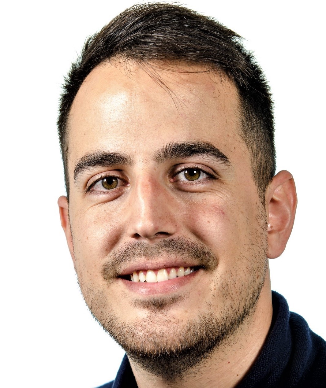
PhD fellow, Dept. of electrical engineering and computer science; Summer school facilitator.
