Mohammed R. S. Sunoqrot - PhD project - NTNU Biotechnology
Computer-Aided Diagnosis of Prostate Cancer Using Multiparametric MRI
Mohammed R. S. Sunoqrot
Research Group: MR Cancer group
Department of Circulation and Medical Imaging, Faculty of Medicine and Health Sciences

In 2017-2021, NTNU Biotechnology funded my PhD project aimed to participate in improving the diagnosis of prostate cancer.
The reason for selecting prostate cancer is that it is the most commonly diagnosed cancer in men and the second leading cause of cancer-related deaths in men worldwide.
In recent years, there have been new advances in technology and diagnostic procedures that have helped improve the diagnosis of prostate cancer and increase patient survival rates. Early and accurate diagnosis of prostate cancer is important for better treatment of the disease. In the last decade, international guidelines for the acquisition and interpretation of prostate cancer images have been introduced to improve the diagnosis of prostate cancer. The guidelines recommend the use of multiparametric magnetic resonance imaging (mpMRI), which captures different imaging modalities during the scan.
To diagnose the patient based on the images, we need to interpret the collected images. Nowadays, this is done qualitatively and manually by radiologists. This approach has a number of limitations, such as the different decisions radiologists make on the same case, the relatively long time it takes to read the images, and the limited number of cases radiologists can handle.
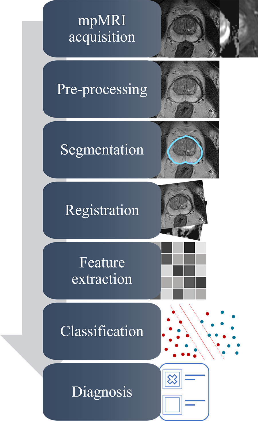
Automated computer-aided detection and diagnosis (CAD) has the potential to overcome manual image reading and leverage mpMRI by implementing quantitative models to automate, standardize, and support reproducible interpretation of radiological images. To provide an efficient and trustworthy diagnosis, each stage of a CAD system should be generalizable, transparent, and robust.
As good as CAD may be, CAD has not yet been integrated into clinical practice. This is mainly because many of CAD systems lack generalizability, transparency, and robustness. To increase trust in CAD, its performance should be improved, controlled, and generalized. Therefore, the aim of this PhD project was to facilitate the integration of automated CAD systems for prostate cancer using mpMRI into clinical practice by developing and evaluating new image normalization, segmentation, and quality control methods to improve the generalizability, transparency, and robustness of the CAD workflow.

During the project lifetime, we developed and introduced two methods: (1) a novel automated method for T2-weighted (T2W) MR image normalization of the prostate, mainly to improve CAD generalizability (2) a fully automated quality control system for deep learning-based segmentation of the prostate on T2W MRI, mainly to improve CAD transparency. We have published both of the methods and we made their code publicly available for anyone to use. In addition, we investigated the reproducibility of the deep learning-based segmentations of the prostate gland and zones to ensure that CAD uses reproducible and robust models.
In summary, this PhD project has developed advanced image pre-processing and quality control methods for CAD of prostate cancer using mpMRI. Ultimately, these automated methods can help improve the performance of CAD and increase confidence in the systems, which is an important step toward their adoption in clinical practice.
Main supervisor: Tone Frost Bathen
Project period: 2017-2021
