Photo-dynamic therapy (PDT) - Biophotonics and Spectroscopy
Photo-dynamic therapy (PDT)
Cancer treatment is usually dependent on agents killing the cancer cells. One method to achieve this is to insert molecules, so called photo-sensitizers (PS), into the relevant cell system or tissue to destroy. Shining light of a selected wavelength, the PS will respond and locally form toxic singlet oxygen and similar reactive oxygen species (ROS) named photodynamic therapy (PDT). In the research we test and develop new PS together with external molecular designers and medical researchers specializing in certain cancers.
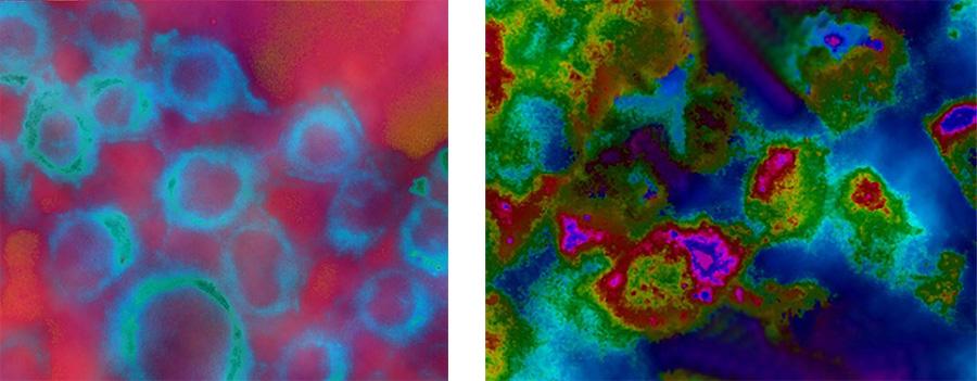
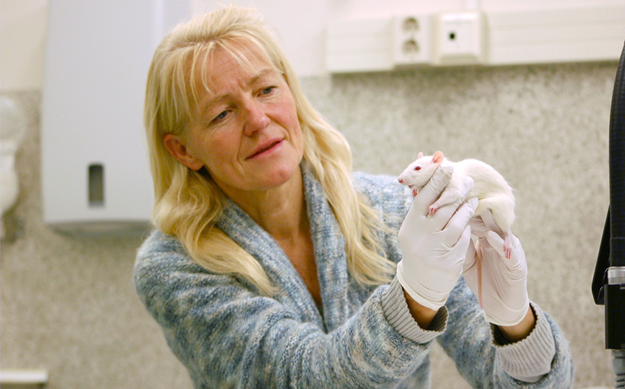
Most of the work is basic research using cell models (in vitro) and established animal models, in vivo, (bladder cancer / gliomas) to explore toxicity and modes of cell death. The activity is tightly connected to the ‘biophotonics lab’ and also use the cell lab and molecular imaging platforms at NTNU/IFY. Fluorescence microscopy studies on human colon carcinoma cells demonstrate remarkable PDT efficiency (Figure 1).

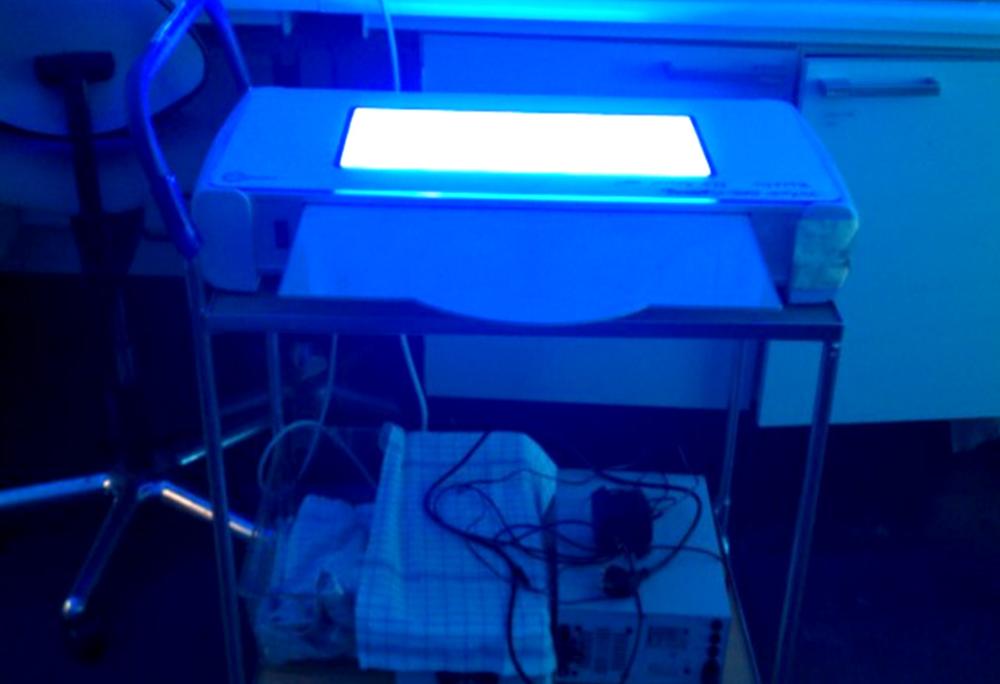
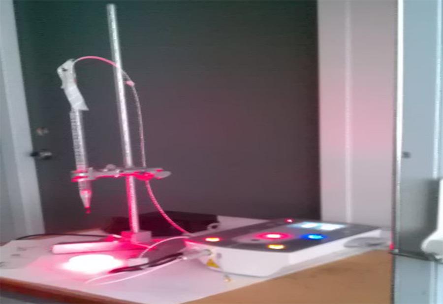
The main interest is to understand mechanisms and development of functional therapies of local tumors using different sensitizers and light sources. Mammalian cells produce light sensitive porphyrins (e.g. protoporphyrin IX) which after stimulating by 5-aminolevulinic acid (ALA), one of the central compounds in heme biosynthetic pathway, results in overproduction of endogen produced porphyrin molecules (ALA-based PDT). Together with light, photochemical reactions destroy cancer cells but not normal cells and this specificity is one of the strongest advantages of PDT. The method is well suited for both treatment and diagnosis of several cancers and diseases. Two available light sources are present in Figure 2 and 3.


The interdisciplinary field of PDT by Gederaas et al. are connected to Dept. of Physics (Prof. Mikael Lindgren and co-workers), Department of Chemistry and Department of Clinical and Molecular Medicine together different Departments at St. Olavs Hospital (Trondheim), UiO and UiT. International researchers from Univ. of Dublin, Univ. of München, Univ. of Porto, Univ. of Lyon, Univ. of Salzburg, Univ. of Sofia, Univ. of Thessaloniki and Univ. of California, are partners in the studies.
Publications
Examples from in vivo projects (bladder cancer model)
- Gederaas O. et al. Photochem. Photobiol. Sci. 2017, 16, 1664-76
- Larsen E. et al. J. Biomed. Optics, 2008,13(4)
- Baglo Y. et al. Photochem. Photobiol. Sci. 2011, 10, 1137-45
- Gederaas O. et al. Peptide. Transl. Oncol. 2014 7(69, 812-23

Examples from ex vivo studies
- Patent: WO 2015/162279 A1. October 29th 2015. Modification of extracorporeal photopheresis technology with porphyrin precursors. International application published under the patent cooperation treaty (PCT). Gederaas is one of 6 inventors.
Examples from in vitro studies
- Lindgren M. et al. Molecules 25, 2020, 1127
- Bogoeva V. et al., Photodiagn. Photodyn. Ther., 2016, 14, 9-17
All informasjon fra Cristin database
Companies that have been parts of ongoing research project in PDT
- PCI Biotech AS, Oslo
- Photocure ASA, Oslo
- APIM Therapeutics, Trondheim
- Byggteknikk Invest AS, Trondheim
