Mass staining and immonohistochemistry – Chemosensory laboratory – Department of psychology
Mass staining and immunohistochemistry

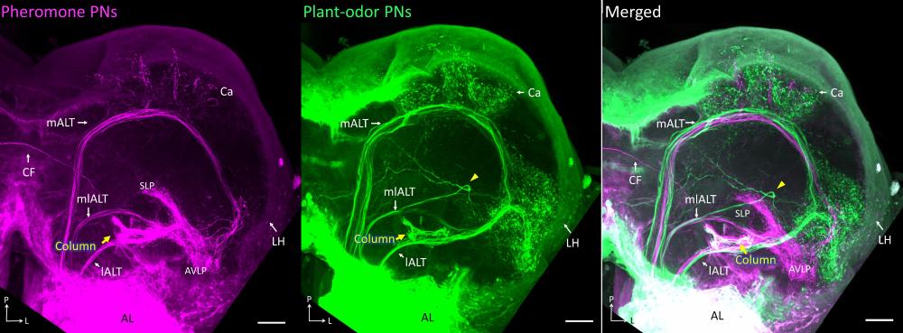
Confocal images of a mass-stained preparation showing the three main antennal-lobe tracts (ALTs), which correspond to the mammalian olfactory tract. Different fluorescent dyes were applied to the male specific antennal-lobe glomeruli (pheromone/magenta) and the ordinary glomeruli (plant odor/green), respectively. White in the merged image to the right, indicates regions receiving overlapping input from MGC and OG. Adapted from Chu et al., 2020, Front. Cell. Neurosci., doi: 10.3389/fncel.2020.00147
Bulk application of fluorescent dyes into an area of interest allows us to visualize, by means of confocal microscopy, the gross morphology of particular neuronal pathways involved in a functional system. By injecting two dyes possessing distinct optical characteristics into different brain areas, we can uncover putative connections between the axon terminals originating from the various regions. The immunostaining technique, on its side, offers the opportunity to stain neuron populations based on the transmitter they utilize (for example GABA and serotonin) or according to the presence of certain molecules, such as synapsin, which is present in all presynaptic terminals.
Lab members
- Xi Chu, Research fellow
- Elena Ian, Research fellow
- Jonas Kymre, Research fellow
- Nicholas Kirkerud, Research fellow
- Mikkel N. Haug, Master student
- Anjela Brianne Griffin, Master student
- Christian Ferdinand Lae Vale, Student at the clinical study program in psychology
- Hanna Mustaparta, Professor emeritus
