X-ray physics
X-ray Physics
The X-ray physics group is Norway’s leading research unit in X-ray physics and imaging. It is associated with the Centre of Excellence (CoE) PoreLab.
Development of experimental methods, instrumentation and applications of advanced X-ray Scattering and Imaging techniques is the core interest of the group.
Our current project portfolio is related mainly to advanced imaging with focus on time-resolution, contrast enhancement, X-ray microscopy and computational methods.
Studies of porous media and mesoscale physics are central, with strong links to geophysics, biophysics and material science.
Computed Tomography

Nikon HT225 microfocus CT-scanner, equipped with a variety of transmission and reflection targets. The CT scanner operates with attenuation contrast, and is versatile operating with samples from a few mm, with nominal resolution ~1-2 μm, up to samples that are 10-15 cm diameter. The setup is equipped with a robust sample stage that can take loads up to 50 kg, allowing for operations with complex sample environments.
If you want to book the tomography lab, use the link below to see when the lab is available. Then make an appointment with the staff engineer.
Tomography lab booking system
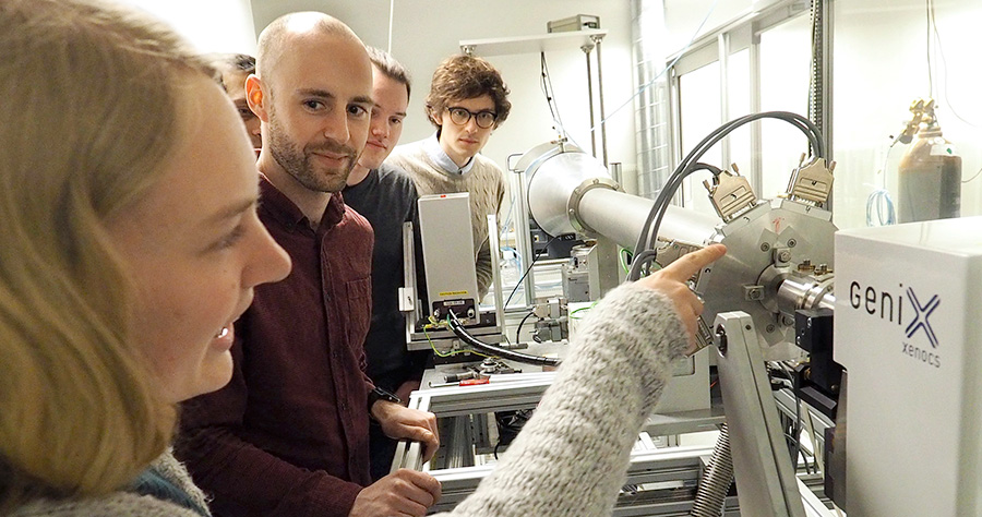
X-ray Diffraction and Scattering

The custom built X-ray scattering setup is equipped with a rotating anode source, Pilatus 1M detector and Huber sample stage . The setup is suitable for both SAXS and WAXS analysis with an opportunity to use a variety of custom built sample environments.
X-ray Radiography

A custom-built setup designed for in situ imaging of evolving microstructures/phase fronts in thin radiographic cells. The system consists of a VISCOM XT9100 microfocus source (Mo, Ag or Cu), and a Vosskuhler CCD-camera, equipped with a SCINT-X pixeled scintillator, optimized for 17 keV radiation. The nominal performance characteristics of the setup is about 4-μm spatial resolution with frame rates up to 6 per second. The facility is unique, with only one similar setup in operation worldwide.
Fourier Microscopy

A custom built optical microscope based on Fourier Ptychography that uses a computational routine to obtain an image of higher resolution than that permitted by the optics. The computationally reconstructed images are quantitative i.e. the phase information is also retrieved. The instrument offers a large field of view and high resolution. The resulting spatial resolution is 700nm and the field of view is 3 X 3mm.
Access to Large Scale Infrastructures
The Research group are frequent users of these synchrotron radiation facilities
- The European Synchrotron (ESRF). The ESRF facility is a joint research facility situated in Grenoble, France.
- Swiss Light Source (SLS). SLS at Paul Scherrer Institut (PSI) is a third-generation synchrotron light source.
- MAX IV Laboratory. Swedish national laboratory providing scientists with the most brilliant X-rays for research.
- Diamond Light Source. The UK's national synchrotron science facility.
Within NTNU the research group have access to:
ICONIC
FRINATEK (Research Council of Norway), 2020-2024
3D- imaging of biomineralisation at the nanoscale using time resolved X-ray imaging and scattering.
Project Leader: Dr. Basab Chattopadhyay (NTNU, Norway)
Consortium Partners: Prof Yves H. Geerts (ULB, Belgium), Dr. Yuriy Chushkin (ESRF, France), Prof. Ragnvald Mathiesen (NTNU), Prof. Dag Breiby (NTNU), Dr. Narayanan Theyencheri (ESRF, France), Prof. Reider Lund (UiO, Norway), Prof. Alain Gibaud (Le Mans University, France).
Poreflow
NANO2021 (Research Council of Norway), 2020-2024
Combined X-ray and neutron tomography for the understanding of flow in porous media
Project Leader: Prof. Dag Breiby (NTNU)
Consortium Partners: Prof. Ragnvald Mathiesen (NTNU), Prof. Francois Renard (UiO), Prof. Erik Johannessen (USN), Prof. Knut E. Aasmundtveit (USN)
4D-CT
FRINATEK(Research Council of Norway), 2018-2022
Computational and experimental framework for time resolved X-ray tomography
Project Leader: Prof. Dag Breiby (NTNU)
Consortium Partners: Prof. M. Nadeem Akram(USN, Norway), Prof. Anne Elster (NTNU), Dr. Andreas Menzel (PSI, Switzerland), Prof. Alain Gibaud (Le Mans University, France)
COMPMIC
NANO2021 (Research Council of Norway), 2018-2021
Quantitative computational optical and X-ray microscopy schemes for monitoring mesostructured and porous objects in real time
Project Leader: Prof. Dag Breiby (NTNU)
Consortium Partners: Prof. M. Nadeem Akram(USN, Norway), Prof. Anne Elster (NTNU), Dr. Andreas Menzel (PSI, Switzerland), Prof. Alain Gibaud (Le Mans University, France)
Cutting Edge
PETROMASK2 (Research Council of Norway), 2018-2021
Advanced methods for analysis of shale cuttings
Supervisor: Prof. Dag Breiby (NTNU)
Project Lead: Dr. Pierre Cerasi , Dr. Cathrine Ringstad, Dr. Malin Torsæter (SINTEF Industry)
Previous projects
- CO2 PLUG (RCN: CLIMIT)
- RECX - National research infrastructure for X-ray scattering and imaging (RCN: INFRASTRUKTUR)
- Towards coherent imaging of working catalyst nanoparticles (RCN: SYNKROTRON)
- ColdWear (RCN: KMB)
- Expériences et simulation de clichés GISAXS de films minces mésoporeux ou fonctionnalisé (RCN: Aurora)
- RAtional DEsign of blends for bulk heterojunction SOLar cells - RADESOL (M-ERA.NET , EU funded research)
Publications of Ragnvald Mathiesen
Publications of Dag W. Breiby
Publications of Basab Chattopadhyay
Master's project
Several Master's projects available
Interested students should get in touch with the PIs.
PhD and postdoc positions are posted on https://www.jobbnorge.no/
Post-doctoral candidates interested in submitting applications for Marie Sklodowska Curie Action (MSCA) Individual Fellowships are invited to contact the PIs directly. The NV-faculty of NTNU provides support for preparation of grant applications through the FINS initiative.
Principal investigators
PhD Candidates
-
Mukul Jaiswal Researcher
mukul.jaiswal@ntnu.no Department of Physics -
Giacomo Luani PhD Candidate
giacomo.luani@ntnu.no Department of Physics -
Arshitha Mathew PhD Candidate
+4740328551 arshitha.mathew@ntnu.no Department of Physics -
Fazel Mirzaei
+4746219422 fazel.mirzaei@ntnu.no -
Soumya Pallipotta PhD Candidate
+4796813237 soumya.pallipotta@ntnu.no Department of Physics -
Shibi Tharayanmaru Palliyalil PhD Candidate
shibi.t.palliyalil@ntnu.no Department of Physics -
Joe Stickland PhD Candidate
joe.stickland@ntnu.no Department of Physics -
Daniyal Younas
daniyal.younas@ntnu.no -
Jessica Zeman PhD Candidate
jessica.zeman@ntnu.no Department of Physics